Zootaxa
2345:
43-59 (2010)
Taxonomy of the sand
bubbler crabs Scopimera globosa De Haan, 1835, and S. tuberculata
Stimpson, 1858 (Crustacea: Decapoda: Dotillidae) in East Asia, with description
of a new species from the Ryukyus, Japan
Kingsley J. H. Wong, Benny K. K. Chan & Hsi-Te Shih*
Sand bubbler crabs of
the genus Scopimera are common on sandy shores in East Asia yet the
taxonomy of the species remains unclear. Scopimera globosa De Haan, 1835,
the type species, was described from Japanese specimens and also occurs in Korea
and China. Scopimera tuberculata Stimpson, 1858, described from Japan,
has been regarded a junior synonym of S. globosa, but the types had long
been lost. Some workers have considered the two taxa distinct and S.
tuberculata has been recorded from South China. In the present study, we
confirm using male gonopod morphology and molecular analysis, that the early
records of S. tuberculata from Hong Kong and S. globosa from
Taiwan are in fact S. intermedia Balss, 1934. The present study regards
S. tuberculata as a subjective junior synonym to S. globosa. A new
species, Scopimera ryukyuensis sp. nov. from the Ryukyus, is identified and
described herein. The new species is close to S. globosa but can be
separated by carapace characters. The mitochondrial cytochrome oxidase I (COI)
gene revealed basepair (bp) difference between the new species and other
Scopimera spp. to be at the interspecific level, at least 28 bp (4.3%).
Key words: Scopimera
ryukyuensis, Dotillidae, cytochrome oxidase I, taxonomy, Ryukyus, Japan.
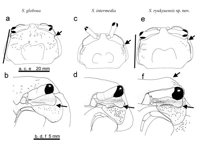
FIGURE 1. Scopimera
globosa De Haan, 1835: dorsal view of carapace (a) and suborbital ridge and
pterygostomian region (b); S. intermedia Balss, 1934: dorsal view of
carapace (c) and suborbital ridge and pterygostomian region (d); S.
ryukyuensis sp. nov.: dorsal view of carapace (e) and suborbital ridge and
pterygostomian region (f). a, c, e: note arrow highlighting relative size of
extra-orbital angle and solid line indicating direction of ridge behind the
extra-orbital angle (which is diagnostic between S. ryukyuensis sp. nov.
and S. globosa). b, d, f: arrow noting raised branchial region viewed
from front in S. ryukyuensis sp. nov. (e) and relative conspicuousness of
sub-orbital ridge: that of S. ryukyuensis sp. nov. being most pronounced.
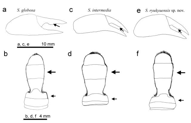
FIGURE 2. Scopimera
globosa De Haan, 1835: chela (a) and male abdomen (b); S. intermedia
Balss, 1934: chela (c) and male abdomen (d); S. ryukyuensis sp. nov.:
chela (e) and male abdomen (f). a, c, e: arrow pointing tooth at inner margin of
movable finger: that of S. intermedia Balss, 1934, being most
rudimentary. b, d, f: large arrows showing dimensions of the sixth abdominal
somite and small arrow indicating the convexity of distal border of the fourth
somite.
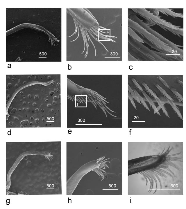
FIGURE 3. Scopimera
globosa De Haan, 1835: G1 entire view (a), tip (b) and magnification of
setae (c); S. intermedia Balss, 1934: G1 entire view (d), tip (e) and
magnification of setae (f); S. ryukyuensis sp. nov.: G1 entire view (g),
tip (h) and tip observed under light microscope (i). Scale bars in μm.
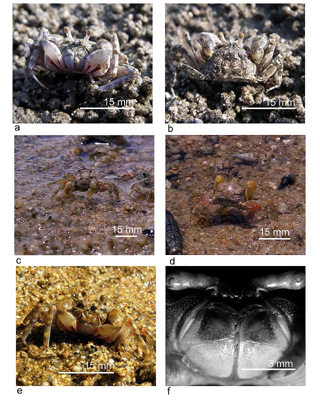
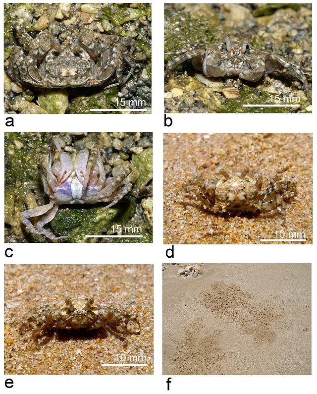
FIGURE 4. Photographs in
life (a–e). Scopimera globosa De Haan, 1835: male frontal view (a) and
dorsal view (b); S. intermedia Balss, 1934: male frontal view (c) and
dorsal view (d); and S. ryukyuensis sp. nov.: male frontal view (e) and close-up
of external maxillipeds from preserved specimens, showing ring-like marking on
merus (f).
FIGURE 5. Photographs in
life Scopimera ryukyuensis sp. nov. (a–e): dorsal view (a), frontal view
(b) and ventral view (c) of male and dorsal view (d) and frontal view (e) of
female. External architecture of burrow at Sedake, Okinawa, the Ryukyus (f).
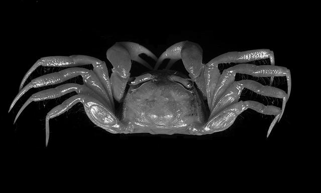
FIGURE 6. Dorsal view of
the neotype of Scopimera tuberculata Stimpson, 1858. A male (cw 9.9 mm,
cl 7.6 mm) (CBM-ZC 4195) deposited in Natural History Museum and Institute,
Chiba, Japan.
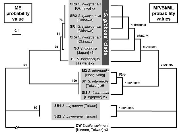
FIGURE 7. A minimum
evolution (ME) tree (left) and maximum parsimony (MP) tree (right) of the
Scopimera species from East Asia and Dotilla, based on 658 basepairs of the
cytochrome oxidase I genes. Probability values at the nodes represent confidence
values for ME (left), MP, Bayesian inference (BI) and maximum likelihood (ML)
(right). For haplotype names see Table 1. Species name and locality are behind
each haplotype name.
 PDF
PDF
Copyright © 2010 Hsi-Te
SHIH